JPT No. 20 – Progressive photonics: methods and applications of sequential imaging using visible and non-visible spectra to enhance data-yield and facilitate forensic interpretation of fossils
Michael J. Eklund1, Arvid K. Aase2, and Christopher J. Bell1
1 – Vertebrate Paleontology Laboratory, Jackson School of Geosciences, The University of Texas at Austin, Austin Texas, 78712 USA
2 – Fossil Butte National Monument, Kemmerer, Wyoming, 83101 USA
ABSTRACT
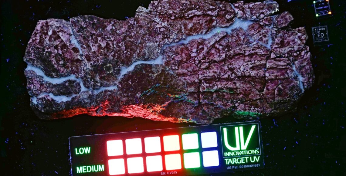
Digital imagery plays a critical role in the documentation and study of fossil specimens. Progressive Photonics is a suite of digital imaging techniques that generate images that yield important and novel insights into the preservation and taphonomic history of fossils, and facilitate rapid comparative review of those images. The techniques are systematic, reproducible, simple, quick, and inexpensive and can be applied successfully in various settings including field sites, laboratories, and museums. We developed, applied, and improved these techniques extensively over the last six years to generate investigative and archival images of paleontological specimens ranging in size from microscopic to very large, from diverse geologic settings, and with taphonomic histories ranging from simple to complex. This imaging strategy uses the visible and invisible portions of the electromagnetic spectrum through utilization of readily available digital cameras and lighting coupled with less commonly used, but easily obtained broad-spectrum UV lighting. This paper demonstrates a standardized, objective sequence of images that can be adapted for various purposes, and that does not rely on subjective image manipulations that commonly are associated with other digital imaging practices. Careful review of the resultant imagery reveals subtle differences that would otherwise remain un-noticed, but contribute important information to the interpretation of a specimen. The sequential imaging process, and the images themselves, promote and enhance scientific investigation and are excellent archival resources for documenting the condition and history of a specimen. The efficacy of Progressive Photonics is demonstrated in three important areas: differentiation of specimen from matrix, differentiation of soft anatomical tissues, and identification of man-made interventions that reflect important aspects of the taphonomic history of specimens.
RESUMO [in Portuguese]
Imagens digitais desempenham um papel fundamental na documentação e estudo de espécimes fósseis. Progressive Photonics é um conjunto de técnicas de imagem digital que gera imagens que revelam importantes e novas perspectivas de preservação e história tafonómica de fósseis, e que facilitam a rápida revisão comparativa das mesmas. As técnicas são sistemáticas, reproduzíveis, simples, rápidas, e baratas e podem ser aplicadas com sucesso em vários contextos, incluindo jazidas, laboratórios e museus. Nós desenvolvemos, aplicámos, e melhorámos estas técnicas extensamente nos últimos seis anos de modo a gerar imagens de arquivo e investigação de espécimes paleontológicos, de tamanho microscópico ao muito grande, de diversos contextos geológicos, e com históricas tafonómicas simples a complexas. Esta estratégia utiliza as porções visíveis e invisíveis do espectro electromagnético através da utilização de câmaras digitais e iluminação facilmente disponíveis emparelhadas com menos comum, mas igualmente fácil de obter luz UV. Este artigo demonstra uma sequência objectiva e padronizada de imagens que podem ser adaptadas para vários propósitos, e que não dependem de manipulação subjectiva de imagens que comummente estão associadas com outras práticas de imagem digital. Revisão cuidada das imagens resultantes revela diferenças subtis que de outra forma passariam despercebidas, mas que contribuem com informação importante de um espécime. O processo de imagens sequenciais, e as próprias imagens, promovem e melhoram a investigação científica e são excelentes recursos de arquivamento para documentar a condição e história de um espécime. A eficácia de Progressive Photonics é demonstrada em três importantes áreas: diferenciação do espécime da matriz, diferenciação de tecido anatómico mole, e identificação de intervenção humana que reflectem aspectos importantes da história tafonómica dos espécimes.

