JPT No. 20 – Progressive photonics: methods and applications of sequential imaging using visible and non-visible spectra to enhance data-yield and facilitate forensic interpretation of fossils

Michael J. Eklund1, Arvid K. Aase2, and Christopher J. Bell1

1 – Vertebrate Paleontology Laboratory, Jackson School of Geosciences, The University of Texas at Austin, Austin Texas, 78712 USA
2 – Fossil Butte National Monument, Kemmerer, Wyoming, 83101 USA

ABSTRACT 

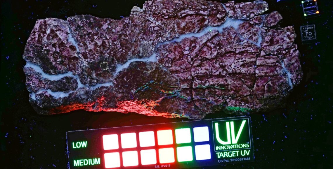
Digital imagery plays a critical role in the documentation and study of fossil specimens. Progressive Photonics is a suite of digital imaging techniques that generate images that yield important and novel insights into the preservation and taphonomic history of fossils, and facilitate rapid comparative review of those images. The techniques are systematic, reproducible, simple, quick, and inexpensive and can be applied successfully in various settings including field sites, laboratories, and museums. We developed, applied, and improved these techniques extensively over the last six years to generate investigative and archival images of paleontological specimens ranging in size from microscopic to very large, from diverse geologic settings, and with taphonomic histories ranging from simple to complex. This imaging strategy uses the visible and invisible portions of the electromagnetic spectrum through utilization of readily available digital cameras and lighting coupled with less commonly used, but easily obtained broad-spectrum UV lighting. This paper demonstrates a standardized, objective sequence of images that can be adapted for various purposes, and that does not rely on subjective image manipulations that commonly are associated with other digital imaging practices. Careful review of the resultant imagery reveals subtle differences that would otherwise remain un-noticed, but contribute important information to the interpretation of a specimen. The sequential imaging process, and the images themselves, promote and enhance scientific investigation and are excellent archival resources for documenting the condition and history of a specimen. The efficacy of Progressive Photonics is demonstrated in three important areas: differentiation of specimen from matrix, differentiation of soft anatomical tissues, and identification of man-made interventions that reflect important aspects of the taphonomic history of specimens.

RESUMO [in Portuguese]

Imagens digitais desempenham um papel fundamental na documentação e estudo de espécimes fósseis. Progressive Photonics é um conjunto de técnicas de imagem digital que gera imagens que revelam importantes e novas perspectivas de preservação e história tafonómica de fósseis, e que facilitam a rápida revisão comparativa das mesmas. As técnicas são sistemáticas, reproduzíveis, simples, rápidas, e baratas e podem ser aplicadas com sucesso em vários contextos, incluindo jazidas, laboratórios e museus. Nós desenvolvemos, aplicámos, e melhorámos estas técnicas extensamente nos últimos seis anos de modo a gerar imagens de arquivo e investigação de espécimes paleontológicos, de tamanho microscópico ao muito grande, de diversos contextos geológicos, e com históricas tafonómicas simples a complexas. Esta estratégia utiliza as porções visíveis e invisíveis do espectro electromagnético através da utilização de câmaras digitais e iluminação facilmente disponíveis emparelhadas com menos comum, mas igualmente fácil de obter luz UV. Este artigo demonstra uma sequência objectiva e padronizada de imagens que podem ser adaptadas para vários propósitos, e que não dependem de manipulação subjectiva de imagens que comummente estão associadas com outras práticas de imagem digital. Revisão cuidada das imagens resultantes revela diferenças subtis que de outra forma passariam despercebidas, mas que contribuem com informação importante de um espécime. O processo de imagens sequenciais, e as próprias imagens, promovem e melhoram a investigação científica e são excelentes recursos de arquivamento para documentar a condição e história de um espécime. A eficácia de Progressive Photonics é demonstrada em três importantes áreas: diferenciação do espécime da matriz, diferenciação de tecido anatómico mole, e identificação de intervenção humana que reflectem aspectos importantes da história tafonómica dos espécimes.

Book Review 2

PREFACE

The current volume of the Journal of Paleontological Techniques is the second number of our yearly series dedicated to the publication of book reviews. We publish reviews every year in February or March, of books that were published the year before, and which cover any aspect related to paleontology or natural history in general.

Deadline for the submissions is generally in mid-January, but proposals will have to be discussed with the editors beforehand, in order to avoid duplicate reviews on the same books. We also invite publishers to notify us about upcoming books, which we will be happy to advertise among our readers and followers on social media.

The current volume includes four book reviews by Abagael R. West, William E. H. Harcourt-Smith, Tom Trapman, and Lukardis C. M. Wencker. The discussed books are about the evolution of horses, humans, invertebrate paleontology, and curators of natural history collections.

PREFÁCIO [in Portuguese]

O volume corrente do Journal of Paleontological Techniques é o segundo número de quatro séries anuais dedicadas à publicação de revisões de livros. Publicamos revisões todos os anos em Fevereiro ou Março, de livros que foram publicados no ano anterior e que digam respeito a qualquer aspecto relacionado com a Paleontologia ou com a História Natural em geral.

O prazo para submissões é, geralmente, meados de Janeiro, mas as propostas têm de ser discutidas com os editores previamente, de modo a evitar revisões duplicadas dos mesmos livros. Convidamos igualmente as editoras a notificar-nos sobre livros que serão publicados em breve, os quais teremos muito prazer em publicitar entre os nossos leitores e seguidores nas redes sociais.

Este volume inclui quatro revisões de livros feitas por Abagael R. West, William E. H. Harcourt-Smith, Tom Trapman e Lukardis C. M. Wencker. Os livros discutidos visam a evolução dos cavalos, a Paleontologia de Invertebrados e os curadores de colecções de História Natural.

JPT No. 19 – A test of a non-consumptive nuclear forensics technique using photon activation analysis of fossils and source matrices

Tyler C. Borgwardt1, Douglas P. Wells1,Darrin C. Pagnac2, Zaijing Sun3, Christian R.Segebade4

1 – Department of Physics, South Dakota School of Mines and Technology, USA

2 – Department of Geology and Geological Engineering, South Dakota School of Mines and Technology, USA

3 – Nuclear Engineering Program, South Carolina State University, USA

4 – Berlin, Germany

Email: tyler.borgwardt@mines.sdsmt.edu

ABSTRACT 

Destructive elemental analysis techniques are common tools in paleontological studies. This study successfully tested the non-destructive technique of photon activation analysis (PAA) for multi-element analysis of fossils and their source matrices. Sample pairs were tested to determine if provenance of fossils could be correlated through their elemental composition. PAA was applied to four pairs of samples, a fossil and a source matrix, each with a different stratigraphic/geographic provenance. Thirty-one total elements were identified: Na, Mg, Ca, Ti, Cr, Mn, Co, Ni, Zn, As, Se, Br, Rb, Sr, Y, Zr, Nb, Mo, Ag, Cd, Sn, Sb, Te, I, Cs, Ba, Ce, Sm, Tl, Pb, U. In each sample 19-26 elements were identified. The lowest concentration detected was 150 ng/g for tellurium. Principal component analysis was applied to the trace elements of the samples. The potential for provenance determination was inconclusive, due to the small number of samples; however, sample pairs showed some separation based on provenance and fossils and matrices were readily distinguishable after analysis.

Keywords: Elemental concentration analysis; photon activation analysis; nuclear forensics; non-destructive; non-invasive; non-consumptive; fossil provenance

RESUMO [in Portuguese]

As técnicas de análises elementares destrutivas são ferramentas comuns nos estudos paleontológicos. Este estudo testou com sucesso a técnica não-destrutiva de análise por activação de fotões (AAF) para análises muti-elementares de fósseis e da suas matrizes rochosas. Pares de amostras foram testados para determinar se a proveniência dos fósseis poderia ser correlacionada através da sua composição elementar. AAF foi aplicada a quatro pares de amostras, um fóssil e a sua rocha matriz, cada um com uma proveniência estratigráfica/geográfica diferente. Foram identificados um total de trinta e um elementos: Na, Mg, Ca, Ti, Cr, Mn, Co, Ni, Zn, As, Se, Br, Rb, Sr, Y, Zr, Nb, Mo, Ag, Cd, Sn, Sb, Te, I, Cs, Ba, Ce, Sm, Tl, Pb, U. Em cada amostra foram identificados 19-26 elementos. A concentração mais baixa detectada foi de 150 ng/g de telúrio. Análise do componente principal foi aplicada aos elementos traço das amostras. O potencial para determinação de proveniência foi inconclusivo devido ao número reduzido de amostras, no entanto, os pares de amostras mostraram alguma separação baseada na sua proveniência e os fósseis e matrizes foram facilmente distinguíveis após as análises.

JPT No. 18 – Coupling focus stacking with photogrammetry to illustrate small fossil teeth

Michael Santella1 & Andrew R. C. Milner1

  1 – St. George Dinosaur Discovery Site at Johnson Farm, St. George, Utah, USA

  Emails: mlsantella@gmail.com (MS), arcmilner@gmail.com (AM)

ABSTRACT 

A macrophotography technique coupled with focus stacking was used to produce source images for photogrammetry of two isolated fossil teeth from the Lower Jurassic Kayenta Formation of southwest Utah. The crown heights of the teeth are approximately 4 mm and 9 mm. The macrophotography was conducted using a Canon digital camera body with a 24-megapixel APS-C sensor. The camera body was attached to a Canon MP-E 65mm lens. A Cognisys focusing rail coupled to a rotation stage was used to manipulate specimens for photography. Focus stacking with Helicon Focus 6 software was used to produce completely focused images for input to Agisoft Photoscan photogrammetry software. The scaled three-dimensional representations created by photogrammetry are available at https://figshare.com/s/d205f5bee8cb2767f902 for specimen UMNH VP 25852 and https://figshare.com/s/af8b4e74db21606a91bd, for specimen UMNH VP 25853. Judging by appearances, retention in the three-dimensional models of shape and surface details from the two-dimensional input images was excellent. A stereo lithography file was also exported for UMNH VP 25852 model, and it was used to produce a 3D-printed model. Regarding identification of the teeth, specimen UMNH VP 25852 has features associated with small theropod dinosaurs. Specimen UMNH VP 25853 has features associated with ornithischian dinosaurs. However, without reference to skeletal remains neither tooth could be unambiguously associated with a specific taxon.

RESUMO [in Portuguese]

Uma técnica de macrofotografia associada ao empilhamento de focagem foi usada para produzir imagens de origem para fotogrametria de dois dentes fósseis isolados, da Formação Kayenta do Jurássico inferior do sudoeste de Utah. A altura das coroas dos dentes é de aproximadamente 4 mm e 9 mm. A macrofotografia foi conduzida usando um corpo de câmara digital Canon, com um sensor APS-C de 24 megapixels. O corpo da câmara foi acoplado a uma lente Canon MP-E 65mm. Um Cognisys com carreto de focagem acoplado a um mecanismo de rotação foi usado para manipular os espécimes para a fotografia. O empilhamento de imagens com diferentes focagem foi realizado com o software Helicon Focus 6 para produção de imagens completamente focadas para serem inseridas no software de fotogrametria Agisoft Photoscan. As representações tridimensionais escalonadas criadas pela fotogrametria estão disponíveis em https://figshare.com/s/d205f5bee8cb2767f902 para o espécime UMNH VP 25852 e https://figshare.com/s/af8b4e74db21606a91bd, para o espécime UMNH VP 25853. Ao que parece, a retenção nos modelos tridimensionais dos detalhes da forma e da superfície das imagens bidimensionais da entrada foi excelente. Um arquivo de litografia estéreo também foi exportado para o modelo UMNH VP 25852, e foi usado para produzir um modelo impresso em 3D. Quanto à identificação dos dentes, o espécime UMNH VP 25852 tem características associadas a pequenos dinossauros terópodes. O espécime UMNH VP 25853 tem características associadas com dinossauros ornitisquios. No entanto, sem associação a um esqueleto permanece ambos os dentes não podem ser inequivocamente associados com um taxon específico.

JPT No. 17 – Scientific illustration and reconstruction of a skull of the diplodocid sauropod dinosaur Galeamopus

Simão Mateus1-5 & Emanuel Tschopp3,4,6

  1 – Museu de História Natural e da Ciência da Universidade do Porto, Porto, Portugal
2 – Departamento de Ciências e Técnicas do Património da Faculdade de Letras da Universidade do Porto (DCTP-FLUP), Porto, Portugal
3 – Museu da Lourinhã, Rua João Luís de Moura, 2530-157 Lourinhã, Portugal
4 – GeoBioTec, Faculdade de Ciências e Tecnologia, Universidade Nova de Lisboa, Caparica, Portugal
5 – Universidade de Évora, Largo dos Colegiais, 7004-516 Évora, Portugal
6 – Dipartimento di Scienze della Terra, Università degli Studi di Torino, Via Valperga Caluso 35, 10125 Torino, Italy

Emails: simaomateus@rocketmail.com (SM), etschopp@unito.it (ET)

ABSTRACT 

High-quality scientific illustration is an important visualization tool for natural sciences. In paleontology, drawings help to guide the reader to important features of the fossils under study, and to remove irrelevant information or strong shadows that might obscure parts of photographs. Furthermore, drawings allow for the deformation of the fossils to be corrected. However, for an accurate interpretation of these reconstruction drawings, it is important to provide a detailed report about the creation of the drawings.
Herein, we describe the methodology of the reconstruction drawing of a skull of the sauropod dinosaur Galeamopus. After preparation and reconstruction of the skull in the laboratory, illustrations were needed to correct natural deformations, restore missing parts, and highlight critical features for anatomical recognition of the several bones. The illustrations were successful thanks to the collaborative work between the paleontologist and the illustrator.

RESUMO [in Portuguese]

Ilustrações científicas de alta qualidade são uma ferramenta importante de visualização nas ciências naturais. Na paleontologia ajudam o leitor a perceber as estruturas anatómicas importantes dos fósseis em estudo, removendo informação irrelevante, ou eliminar zonas escuras que escondam pormenores dos ossos nas fotografias. Além disso, as ilustrações permitem corrigir de ossos deformados. Para a correcta interpretação das reconstruções efectuadas, é importante existirem relatórios detalhados do processo da ilustração.
Vimos descrever a metodologia de ilustração de um crânio de dinossauro saurópode Galeamopus que foi reconstruído. Após a preparação e montagem do crânio no laboratório, as ilustrações tiveram de reajustar as deformações naturais, repor partes em falta, e realçar características essenciais necessárias à compreensão dos diversos ossos. As ilustrações são bem sucedidas graças à colaboração entre o paleontólogo e o ilustrador.

Book Review 1

PREFACE

The current volume of the Journal of Paleontological Techniques is the first number of a new, yearly series dedicated to the publication of book reviews. We aim to publish reviews every year in February or March, of books that were published the year before, and which cover any aspect related to paleontology.

Deadline for the submissions will be December 15, but proposals will have to be discussed with the editors beforehand, in order to avoid duplicate reviews on the same books. We also invite publishers to notify us about upcoming books, which we will be happy to advertise among our readers and followers on social media.

The current volume includes three book reviews by Christine Böhmer, Femke Holwerda, and Emanuel Tschopp. The discussed books are about prehistoric mammals, dinosaurs, and the study of species in a paleontological context.

PREFÁCIO [in Portuguese]

O presente volume do Journal of Paleontological Techniques representa o primeiro número de uma série dedicada à publicação de recensões críticas de livros. Teremos como objectivo a publicação anual de recensões críticas em Fevereiro ou Março de livros que foram publicados no ano precedente e que cubram aspectos relacionados com paleontologia.

As submissões devem ser enviadas até 15 de Dezembro. No entanto, quaisquer propostas devem ser discutidas com os editores antecipadamente de forma a evitar duplicação de trabalhos sobre as mesmas obras. Gostaríamos de convidar as editoras para que nos notifiquem sobre os livros que serão publicados para que possamos divulgá-los junto dos nossos leitores e seguidores nas redes sociais.

O presente volume inclui três recensões críticas escritas por Christine Böhmer, Femke Holwerda, e Emanuel Tschopp. Os livros agora revistos debruçam-se sobre mamíferos pré-históricos, dinossauros e sobre o estudo das espécies em contexto paleontológico.

JPT No. 16 – Preparation of fossil and osteological 3d-printable models from freely available CT-scan movies

Thomas A. Hegna1 & Robert E. Johnson1

1 – Department of Geology, Western Illinois University, Macomb, IL, 61455, USA

Emails: Ta-hegna@wiu.edu, Re-Johnson2@wiu.edu

ABSTRACT

Changes in the online availability of 3D models of skeletal elements and 3D printer technology have made it feasible to develop a fossil or osteological collection from scratch. Many online sites have models that can be freely downloaded and 3D printed. Other sites have CT-scan movies online—which can be downloaded and turned into printable 3D models by using the free software suite SPIERS. Post-processing of the printed models (sanding, painting, etc.) can make them almost visually indistinguishable from real specimens.

RESUMO [in Portuguese]

Alterações na disponibilidade on-line de modelos 3D de elementos esqueléticos e a tecnologia de impressão 3D tornaram possível desenvolver uma colecção, a partir do zero, de osteologia ou fósseis. Muitos sítios on-line têm modelos que podem ser descarregados gratuitamente e impressos em 3D. Outros sítios disponibilizam filmes de tomografias computorizadas que podem ser descarregados e convertidos em modelos 3D para impressão usando programa informático livre SPIERS. O pós-processamento dos modelos impressos (lixamento, pintura, etc.) pode torná-los quase indistinguíveis dos espécimes reais.

JPT No. 14 – Photogrammetry vs. micro-CT scanning for 3D surface generation of a typical vertebrate fossil – a case study

Julia M. Fahlke1 and Marijke Autenrieth1 

1 – Museum für Naturkunde, Leibniz-Institut für Evolutions- und Biodiversitätsforschung, Invalidenstraße 43, 10115 Berlin, Germany

E-mail: Julia.Fahlke@mfn-berlin.de

ABSTRACT

Over the recent years, three-dimensional (3D) surface digitization of fossils has achieved wide application in vertebrate paleontology, be it for reconstruction, morphometric or preservation purposes. The wide array of techniques available for 3D surface generation can, however, be somewhat confusing. Therefore, the aim of our study is to help the paleontologist reach a well-informed decision on which technique to use for standard purposes, taking into account aspects such as accuracy, reproducibility, and efficiency. In this study, we are comparing the above aspects when applying micro-computed tomography (micro-CT), manual photogrammetry and automated photogrammetry to an object typically digitized in vertebrate paleontology: a medium-sized fossil mammal cranium (that of an early Oligocene anthracotheriid). Our results show that manual photogrammetry has a high degree of reproducibility and is the most efficient, least costly method of the ones tested, although more training is required for the unexperienced researcher. Also, when attention is paid to proper lighting and overlap, fewer photographs do not necessarily yield inaccurate results, increasing the speed of data acquisition even more. Disadvantages of CT scanning in external surface generation include the lack of a photo surface, and long post-processing times due to removal of internal surface structures that are not used for this purpose. The technology used for automated photogrammetry, MDS Witikon, offers fast and convenient digitization. Large amounts of data and detail produced, however, may be very useful for the creation of object panoramas but exceed the limits for common use of 3D surfaces.

RESUMO [in Portuguese]

Recentemente, a digitalização da superfície de fósseis a três dimensões (3D) tem tido uma ampla aplicação na paleontologia de vertebrados, quer para propósitos de reconstrução, morfometria ou preservação. O vasto espectro de técnicas disponíveis para a geração de superfícies 3D pode, no entanto, ser confusa. Portanto, o objectivo deste estudo é ajudar o paleontólogo a chegar a uma decisão bem informada sobre que técnica usar para propósitos normais, tendo em consideração a precisão, reproducibilidade e eficiência. Neste estudo, comparamos os aspectos acima quando usando tomografia computorizada, fotogrametria manual e fotogrametria automática para um objecto tipicamente digitalizado em paleontologia de vertebrados: um crânio de um mamífero fossilizado (Anthracotheridae do Oligocénico inferior). Os nossos resultados demonstram que a fotogrametria manual tem um elevado grau de reproducibilidade, é mais eficiente e menos caro dos métodos testados, apesar de requerer mais treino para um investigador com pouca experiência. Além disso, quando a atenção devida é tomada à luz e sobreposição, menos fotografias não implicam piores resultados, o que aumenta a velocidade de aquisição. As desvantagens da tomografia computorizada para fins de geração de modelos da superfície externa incluem a ausência de uma foto de superfície bem como um pós-processamento demorado devido à remoção das estruturas internas desnecessárias. A tecnologia usada para fotogrametria automática, MDS Witikon, oferece uma digitalização rápida e conveniente. Grandes quantidades de informação e detalhe são produzidos, contudo, apesar de ser útil para a criação de object panoramas mas excede os limites para o uso comum de superfícies 3D.

JPT No. 15 Special Volume – Symposium Volume of the 2nd Internacional conservation symposium-workshop

Conveners
Sandra Val & Rubén García; Steven J. Jabo; Chris Collis; Vicen Carrió

Edited by
Emanuel Tschopp, Femke Holwerda & Sandra Val

Additional Scientific Editors
Koen Stein, Carla Alexandra Tomás, Ricardo Araújo, Octávio Mateus, Rui Castanhinha, Ángel Blanco, Christine Böhmer, Ignacio Escapa, Matteo Belvedere & João Russo

Technical Editors
Silvia Costa, Emanuel Tschopp, Femke Holwerda

PREFACE

This special volume collects the work of some of the attendees and the international guests to the 2nd International Conservation Symposium-Workshop in Natural History Collections, organized by the CRIP (Centre of Paleontological Restoration and Interpretation) of Els Hostalets of Pierola, Barcelona (Spain), together with the Museum of Natural History of the Smithsonian Institution of Washington (USA), the National Museums of Scotland in Edinburg (UK) and the Natural History Museum of London (UK). This international symposium aimed to create a workspace and study around the concepts of protection and conservation of Natural History Collections, deepening the knowledge and analysis of the different working methods used in this type of collections. Participation in the symposium was a great success with over 60 participants from different parts of the world: Norway, Denmark, Sweden, Israel, England, Portugal, USA and Spain. This second international meeting was a great space for discussion and exchange of experiences, very dynamic and profitable, where professionals from around the world debated and reflected around the conservation, preparation and restoration of Natural History Collections, establishing ties of professional and institutional collaborations among the participants. The symposium organizing committee wishes to thank all participants and collaborating institutions whose participation has made this meeting possible. And also we want to thank the Journal of Paleontological Techniques for the realization of the Symposium Volume. The team of the Journal of Paleontological Techniques put a lot of effort in editing and publishing this volume monograph.

Fossil tracks and trackways: the dilemmas of preservation
Neville Agnew & Martha Demas  (pdf)
 

Scanning Electron Microscope (SEM) shows undesirable effects of ultrasonic cleaning on recent mollusk shells

Marina Rull, Marta Pérez & Francesc Uribe (pdf)

Study on the physical-mechanical comportment in six gap fillers for fossil reintegration

Tatiana Martínez Riera & Begoña Carrascosa Moliner (pdf)

Consolidation of bone material: chromatic evolution of resins after UV accelerated aging

M. Amparo Linares Soriano & M. Begoña Carrascosa Moliner (pdf)

Conservation-restoration applied to a fossil adhered to a speleothem (Middle Pleistocene) from Postes Cave (Fuentes de León, Spain)

Maria Cruz Ortega Martínez, Monica Villalba de Alvarado, Hipolito Collado Giraldo, José Ramón Bello Rodrigo, Isabel Domínguez García, Luis F. Nobre da Silva, Ángel Carmelo Domínguez García, Lázaro Rodríguez Dorado, José Manuel Torrado Cárdeno, Jairo González Márquez, Elena García Domínguez & Elena Garrido Fernández (pdf)

In situ conservation strategies at the Pleistocene sites of Pinilla del Valle, Madrid (Spain)

Maria Cruz Ortega, Olalla Teresa Canseco, Ana Pastor, Juan Luis Arsuaga, Alfredo Pérez-González, Cesar Laplana, Belen Márquez & Enrique Baquedano(pdf)

Treating a historic restoration of a Megatherium sp. tail from the Rodrigo Botet Collection in the Museum of Natural Sciences of Valencia

Ester Antón, Begoña Carrascosa, Margarita Belinchón & Sandra Illobre (pdf)

Past preparation procedures and contemporary conservation techniques applied on a holotype (Museo Geominero, Madrid, Spain)

Eleuterio Baeza, Silvia Menéndez, Ana María Bravo & Laura Ruiz (pdf)

Unfavorable microclimate conditions in exhibition rooms: early detection, risk identification and preventive conservation measures

Dario Camuffo & Chiara Bertolin (pdf)

JPT No. 13 Special Volume – Symposium Volume of the 1st Internacional conservation symposium-workshop

Conveners
Sandra Val, Steven J. Jabo & Vincen Carrió

Edited by
Emanuel Tschopp & Sandra Val

Additional Scientific Editors
Koen Stein, Carla Alexandra Tomás, Ricardo Araújo, Octávio Mateus, Peter Falkingham & Rui Castanhinha

Technical Editors
Silvia Costa, Emanuel Tschopp, Femke Holwerda

PREFACE

This special volume collects the work of some of the attendees and the international guests to the 1st International Conservation Symposium-Workshop in Natural History Collections, organized by the CRIP (Centre of Paleontological Restoration and Interpretation) of Els Hostalets of Pierola, Barcelona (Spain), together with the Museum of Natural History of the Smithsonian Institution of Washington (USA) and the National Museums of Scotland in Edinburg (UK). This international symposium aimed to create a workspace and study around the concepts of protection and conservation of Natural History Collections, deepening the knowledge and analysis of the different working methods used in this type of collections. Participation in the symposium was a great success with over 80 participants from different parts of the world: Norway, Denmark, Sweden, Israel, Germany, England, Portugal, USA, Australia and Spain. This first international meeting was a great space for discussion and exchange of experiences, very dynamic and profitable, where professionals from around the world debated and reflected around the conservation, preparation and restoration of Natural History Collections, establishing ties of professional and institutional collaborations among the participants. The symposium organizing committee wishes to thank all participants and collaborating institutions whose participation has made this meeting possible. And also we want to thank the Journal of Paleontological Techniques for the realization of the Symposium Volume. The team of the Journal of Paleontological Techniques put a lot of effort in editing and publishing this volume monograph.

In situ conservation strategies: a case study of archeopaleontological remains from the Early Pleistocene site of El-Kherba (Ain Hanech), Algeria

Elena Lacasa-Marquina, Pilar Fernandez-Colón, Aldjia Hamlat, Latefa Marouf, Zoheir Harichane & Mohamed Sahnouni  (pdf)

Bones and sediments: part of a synergetic continuum

Gail Gali Beiner & Rivka Rabinovich (pdf)

Preliminary results on the chemical preparation of dinosaur eggshells

Sandra Val, Rubén García & Domingo López  (pdf)

Preparation of a turtle fossil from the Pliocene site of Camp dels Ninots (Caldes de Malavella, Girona, Spain)
Souhila Roubach, Bruno Gómez de Soler, Gerard Campeny Vall-Llosera & Juan Ignacio Morales (pdf)

The Bristol Dinosaur Project – a conservation and preparation overview

Pedro A. Viegas & Michael J. Benton (pdf)

Two examples of preventive conservation actions in the Museu de Ciencies Naturals de Barcelona (MCNB): inspection of specimens and substitution of packaging

Maria Vila, Marta Pérez, Olga Muñoz & Eulàlia Garcia-Franquesa (pdf)

Restauration of mounted animals – new techniques in old taxidermies

Angel Blanco & Gema Solis (pdf)

Procedures and materials used in the mounting of two birds which belong to the Natural Sciences National Museum (MNCN-CSIC) and the Complutense University of Madrid (UCM)

Rita Gil Macarrón, Sonia Santos Gómez, Luis Castelo Sardina, Margarita San Andrés Moya & Andrés Sánchez Ledesma (pdf)

Pollutants in the museum environment – minimizing damage in storage and display

Pamela Hatchfield  (pdf)

Exploring the common ground between organic artifacts and natural history specimens: we share problems – can we share solutions?

Allyson Rae (pdf)

The collections of vertebrates of the Estación Biológica de Doñana (CSIC). Origin and evolution

Teresa García-Díez, M. Rosario Sempere, José Cabot, Javier Juste & Carlos Ibáñez  (pdf)