JPT No. 12 – Photogrammetry in paleontology – a practical guide

Heinrich Mallison1 & Oliver Wings1,2

1-  Museum für Naturkunde Berlin, Invalidenstraße 43, 10115 Berlin, Germany.

2-  Niedersächsisches Landesmuseum Hannover, Willy-Brandt-Allee 5, 30169 Hannover, Germany.

Emails: heinrich.mallison@gmail.com (HM), dr.wings@gmail.com (OW)

ABSTRACT

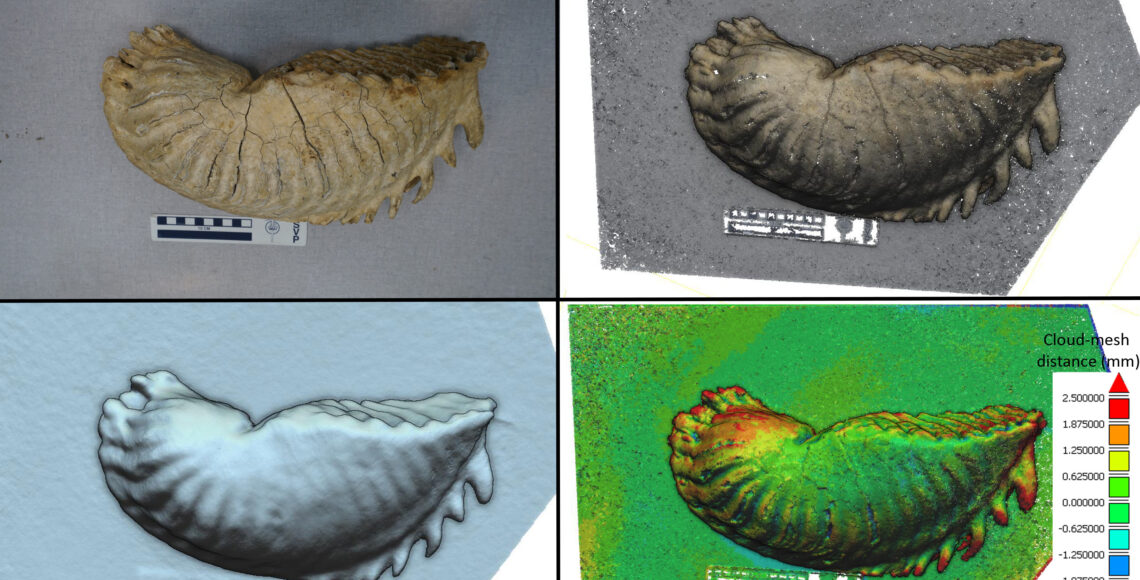
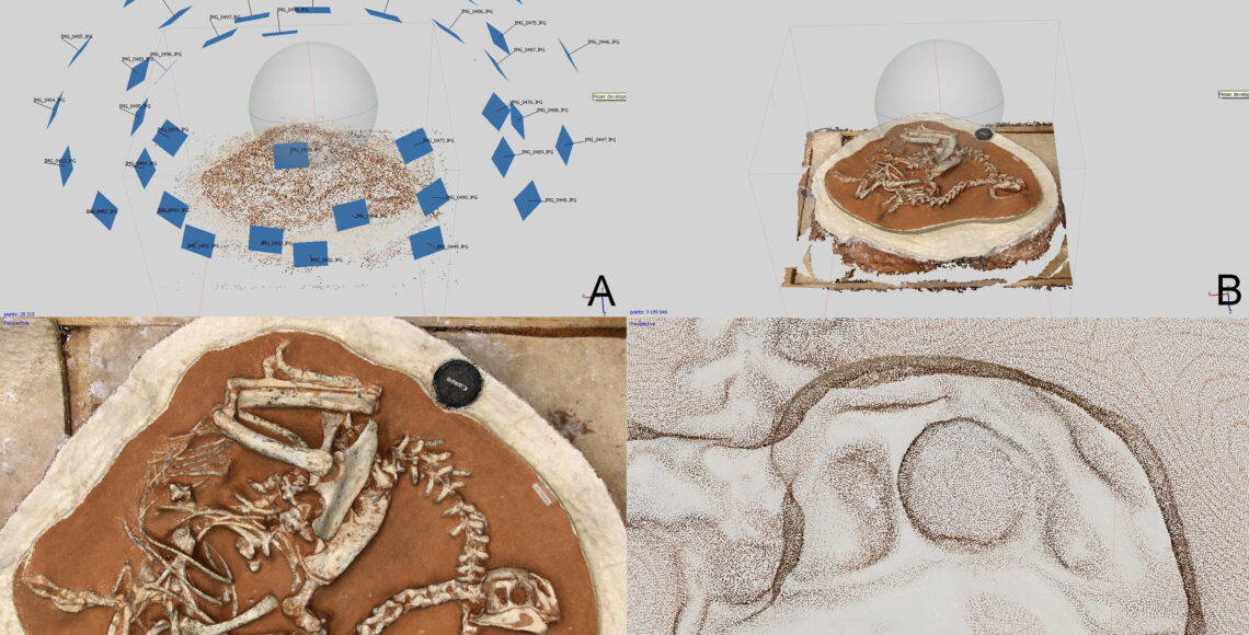
Photogrammetry is increasingly becoming the gold standard for surface digitizing in paleontology. We present techniques for specimen handling, photography and image handling in photogrammetry software that are specially adapted to typical use cases in paleontology, but are also applicable in other science disciplines like archaeology and art history.

RESUMO [in Portuguese]

A fotogrametria está a tornar-se a técnica standard para a digitalização de superfícies em paleontologia. Apresentamos várias técnicas para o manejamento de espécimes, fotografia e processamento de software de imagem que está especialmente adaptado a casos típicos em paleontologia, mas é também aplicável noutras disciplinas científicas como a arqueologia e história de arte.

JPT No. 11 – Low cost 3D scanning using off-the-shelf video gaming peripherals

Peter L. Falkingham1

1- Department of Comparative Biomedical Sciences, Structure & Motion Laboratory, Royal Veterinary College, London, UK and Department of Ecology Evolutionary Biology, Division of Biology and Medicine, Brown University, USA Email: pfalkingham@rvc.ac.uk

ABSTRACT 

Digitization of specimens is becoming an ever more important part of palaeontology, both for archival and research purposes. The advent of mainstream hardware containing depth sensors and RGB cameras, used primarily for interacting with video games, in conjunction with an open platform used by developers, has led to an abundance of highly affordable technology with which to digitize specimens.  Here, the Microsoft® Kinect™ is used to digitize specimens of varying sizes in order to demonstrate the potential applications of the technology to palaeontologists. The resulting digital models are compared with models produced using photogrammetry. Although the Kinect™ generally records morphology at a lower resolution, and thus captures less detail than photogrammetric techniques, it offers advantages in speed of data acquisition, and generation of a completed mesh in real time at the point of data collection. Whilst it is therefore limited in archival applications, the ease of use and low cost, driven by strong market competition, make this technology an enticing alternative for studies where rapid digitization of general morphology is desired.

Keywords:     Scanning; 3D; digital; model; laser; Kinect; digitization, photogrammetry

INTRODUCTION

The ability to digitally capture the 3D morphology of a specimen has revolutionised palaeontology over recent years. Working in the virtual realm permits investigators to section, profile, maniulate and colour a specimen in ways that would otherwise be difficult or impossible, and often destructive, if applied to the real fossils. The use of digital models has facilitated a wide range of research into areas including locomotion, feeding, body mass calculations, documentation, conservation, hydro- and aerodynamics, and many others (Anderson et al., 2011Bates et al., 2012Bates and Falkingham, 2012Bates et al., 2009Falkingham et al., 2009Farlow et al., 2012Gidmark et al., 2013Hutchinson et al., 2011Panagiotopoulou et al., 2011Rayfield, 2007Sellers et al., 2012). Equally importantly, digitization of specimens in the internet age has enabled an unprecedented level of data sharing and collaboration, exemplified by online repositories such as Digimorph (www.digimorph.org), and online journals such as this, which enable digital models to accompany publications as electronic supplementary material (see appendices).

Until recently, however, digitization of fossils remained the purview of those with access to expensive hardware such as computed tomography (CT) and laser scanners, or expensive software-based photogrammetric solutions. While CT machines remain a requirement for internal morphology, methods with which to digitize external morphology have reached such a low cost that they have become available to anyone. Photogrammetric techniques can now be employed with a basic consumer camera, desktop PC, and free software (Falkingham, 2012).

One of the major developments in photogrammetric software most recently has been incorporating the GPU, or Graphics Processing Unit, found in many modern desktop and laptop computers – particularly in machines built for running computer games. The GPU contains many cores, far exceeding the number of cores found on the CPU, or Central Processing Unit (the ‘processor’ of the computer). While GPU cores are more specialized than those on the CPU, they can be used to dramatically speed up 3D applications (providing the software is written to take advantage of the GPU). Importantly, unlike more industrial hardware such as laser scanners, GPUs form part of a major consumer driven market – the video game industry – which drives prices down and processing power up at exceptional rates. Coupled with the falling cost of rapid-prototype machines (3D printers), there is now also a growing consumer demand for 3D digitization, which means that palaeontologists can take advantage of software and hardware developments at prices determined by aggressive market forces.

One such recent development is in using depth sensors, designed for interacting with computer games without a traditional controller, to scan and digitize objects and environments. Such sensors first came into being (at least in the mainstream) with the Microsoft KinectTM for Xbox 360®, released for the gaming console in 2010. This was later followed by the Microsoft KinectTM for Windows®, and the Asus® Xtion Pro, which possess similar specifications but are designed for developers and a personal computer environment.

Although developed for computer games, the low cost depth sensor was quickly ‘hacked’ for other uses, particularly in the fields of robotics (Stowers et al., 2011) and rehabilitation (Chang et al., 2011). Of pertinence here is that one of these uses included real time mapping of the environment, and tracking of the sensor (Newcombe et al., 2011). Despite initially being developed for robots navigating environments, the 3D mapping functionality can be used to create digital 3D models of objects or environments, and software applications designed for this purpose are now available (Izadi et al., 2011). These applications are possible because of the recent increases in consumer GPUs; being able to record a point cloud from the depth sensor, and in real time mesh that point cloud to produce a 3D model.

In this paper, I aim to demonstrate the applicability of these gaming peripherals and associated software packages for scanning and digitising palaeontological specimens.

JPT No. 10 – 3-Dimensional reproduction techniques to preserve and spread paleontological material – a case study with a diplodocid sauropod neck

Tschopp, Emanuel1,2 and Dzemski, Gordon3

1- CICEGe, Faculdade de Ciências e Tecnologia, FCT, Universidade Nova de Lisboa, 2829-516 Caparica, Portugal
2- Museu da Lourinhã, Rua João Luis de Moura, 2530-158 Lourinhã, Portugal.
3- Institut für Biologie und Ihre Didaktik, Universität Flensburg, Germany.

ABSTRACT 

The protection and preservation of irrecoverable, sensitive and fragile objects in museums, exhibitions and collections is critical in various research fields like cultural heritage, human sciences and paleontology. Lately, digitization of such endangered specimens proved to be a valuable tool to convert the objects into a digital form. However, in order to exploit all possibilities that such data could provide for educational and research purposes, it can be useful to transform the digital material back into a physical form. In this case study, a neck of a diplodocid sauropod dinosaur was digitally reproduced by 3D printing (a variant of rapid prototyping). The process is described in detail, and compared to the more classical reproduction using CNC-mills.  CNC-milling is an inexpensive and accurate reproduction technology for large objects, and especially well-designed for producing durable casts. On the other hand, 3D printing is highly accurate to create small or complex objects, but is more expensive and yields more fragile physical objects. As accuracy of the complex shapes of the diplodocid cervical vertebrae was required in order to use them for research, 3D printing was preferred over CNC-milling in this case.

INTRODUCTION

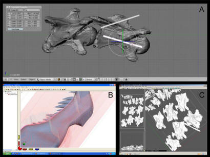
Access to original material or specimens is limited for palaeontologists due to a number of reasons. Fossil material is rare and difficult to access (e.g. distance, museum policies, intense scientific interest). Fossil bones can be heavy or fragile to transport, or the desired element is mounted and on public display in a museum. Three-dimensional reproduction techniques therefore offer a great solution for archiving significant data of paleontological objects (Breithaupt et al., 2004; Remondino et al., 2005). Whereas in the early years of paleontology, physical casting of fossils has proved a valuable alternative way for researchers to undertake their investigations, as casts are usually lighter, less fragile, and easier to handle than original material, in the last 15 to 20 years alternative technologies have been developed to produce digital copies of original specimens (see Zollikofer and Ponce de León, 1995; Chapman et al., 1998; or Andersen et al., 2001 for some of the earliest attempts in Paleontology). Laser, x-ray, or magnet scanner, mechanical digitizers and photogrammetry methods transform matter into bits and bytes to create, manipulate, recreate and change objects assisted by a computer with editing software. For paleontological purposes the digital models can be stored and used for scientific research, spread via internet, or can be displayed virtually in museum exhibitions (Johnston et al., 2004; Mallison et al., 2009). Entire specimens can be measured providing accurate results for further research (Deck et al., 2004). Using simulation methods, bones can be articulated with each other without having to handle them manually – and thus without subjecting them to possible abrasion or damage due to real bone to bone contact (Chapman et al., 2001). They can be subjected to retrodeformation methods in order to re-establish a shape that is supposed to be closer to the original morphology (Motani et al., 2005; Kahzdan et al., 2009; Tschopp and Dzemski, in review now). Moreover, digital models of skeletal elements can be virtually connected with tendons and muscles to reconstruct the entire animal and to study in vivo motion patterns and biomechanical hypotheses (Walters et al., 2001; Bimber et al., 2002; Dzemski and Christian, 2007, 2010).

On the other hand, scientists often stress the importance of studying three-dimensional physical objects to manipulate them by hand and realize the dimensions of the dynamic elements. Physical models can furthermore be used for education and in museum exhibits, and have a great potential to indirectly protect the original objects (Mallison, 2007; Remondino, 2007; Schlader et al., 2007). Instead of the traditional casting process described above, there are now several ways to transform a digital object to a physical model. They can be subdivided into additive (layer by layer “printing”) and subtractive (e.g. carving, milling) techniques.  The additive method used for this case study (3D printing), as well as CNC-milling as example for a subtractive technique will herein be described, and their usage, advantages, and possibilities will be compared and discussed regarding the reproduction of the neck of the diplodocid sauropod SMA 0004 for research purposes.

Abbreviations: CAD: Computer Aided Design; CAM: Computer Aided Manufacturing; CNC: Computer Numerical Controlled; SMA: Sauriermuseum Aathal, Switzerland; STL: Standard Triangulation Language (a file format for 3D models).

JPT No. 9 – How to mount an inexpensive sieving lab

Ricardo Araújo1,2, Carlos Natário1, Matthew Pound3,4

1 – Museu da Lourinhã, Rua João de Luís de Moura, 95, 2530-158 Lourinhã, Portugal.
2 – Huffington Department of Earth Sciences, Southern Methodist University, PO Box 750395, Dallas, Texas, 75275-0395, USA.
3 – School of Earth and Environment, University of Leeds, Leeds, LS2 9JT, United Kingdom.
4 – British Geological Survey, Keyworth, Nottingham, NG12 5GG, United Kingdom.

ABSTRACT 

We present a new sieving technique designed to recover microfossils from mudstones, clays and poorly consolidated sediments. This new technique is designed to be inexpensive, use the minimal amounts of chemicals and recover well preserved microfossils. The inexpensive nature of this methodology makes it suitable for reconnaissance studies, where finances may be limited. Not using large amounts of chemicals helps to protect the fossils and the environment. Investigations on the Jurassic Lourinhã Formation, Portugal yielded a diverse microfossil assemblage including; archosaur teeth, lizard jaws, amphibian jaws, fish remains, ostracods and charcoal. Such a diverse fossil recovery shows the technique is suitable for painstaking palaeoecological studies as well as reconnaissance work.

INTRODUCTION

The use of confocal laser scanning microscopy (CLSM) for the study of inclusions in amber is a recent development. Böker & Brocksch (2002) produced a series of images of insects in Baltic amber using CLSM and identified the potential for 3D imaging of minute detail of taxonomically important morphological structures such as the mandibles and genital organs. Since then, however, very little has been published on CLSM analysis of amber inclusions despite the apparent benefits.

Sieving methods for microvertebrate recovery have rarely (Mateus et al. 1997) been applied to the Lourinhã Formation. The Lourinhã Formation is comprised of fossiliferous fluviodeltaic deposits that outcrop extensively in the Lusitanian Basin, Western Portugal. This formation is mainly composed of intercalated sandstone channels with extensive alluvial mudstone layers (Hill 1989). Within the extensive mudstone layers most fossils including many large vertebrates (Antunes and Mateus, 2003) and microfossils are found (e.g. Ramalho 1967). Microvertebrate fossil faunas have been collected for many years in the Lourinhã Formation, mainly by surface collecting. However, in 2008 the Museum of Lourinhã started a systematic sieving campaign to better understand the overall fauna of the Lourinhã Formation. 

Some of the first microvertebrate finds in Portugal were discovered in the Guimarota mine in 1960 by palaeontologists from the Freilicht Universitat, Berlin (Krebs, 2000). The first account of sieving methods being used in Portugal were published by Kühne (1968), using a constant flow of water and sieves incorportated on a metal barrel. Sieving was applied at the Paimogo theropod embryo nest site (Mateus et al. 1997) from 1994 to 1996, in the search for embryo bones and eggshells (Mateus, 1998) and was also occasionally applied to the Porto das Barcas fossil site, but without much success.

Precise geographical information of sieving sites in the Lourinhã Formation was acquired using GPS coordinates, stratigraphic information and a measured section was acquired by plotting the site photograph with detailed geologic annotation. Samples were taken using a pick axe at 30 – 40cm stratigraphic intervals and within 1 – 2m horizontal spacing. This systematic sieving campaign, applied to the Lourinhã Formation for the very first time, is being used to investigate and assess the composition and diversity of the microfossil fauna.

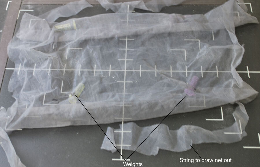
The first use of sieving in the search of microvertebrate remains was performed ca. 1847 by Plieninger, in Germany (McKenna et al. 1994). Early methods of sieving were also used by Moore in England (1867) and later by Wortman and Brown in the United States (1891).  Sieving became well implemented in the palaeontological community after Hibbard (1949) reported using the technique to collect Cenozoic mammal fossils from an unconsolidated sandstone matrix (McKenna et al., 1994). Hibbard introduced the use of screen boxes (wooden boxes with a brass mesh). However, this method requires having water near the work site, is laborious, and requires a large staff (Ward 1984). McKenna (1962; 1965) provided further insight into the screen box technique and proposed a standard model using manufactured rectangular wooden boxes and a regular size steel mesh. Both McKenna’s and Hibbard’s techniques are field-oriented and require the presence of nearby water. McKenna simply optimized Hibbard’s technique by processing a larger quantity of matrix using more screen boxes (almost 300, compared to Hibbard’s dozen), and adapting the method to the constraints of particular sites. Grady (1979) described a new method using mosquito nets instead of the classical material of screen boxes with brass mesh, providing a more field-oriented  method  with easily transported equipment (Ward, 1984).

In contrast, the method reported in this paper is similar to other laboratory-oriented techniques. Described in further detail by Kühne (1971) and Krebs (2000), the “Henkel technique” (Henkel, 1966) is a laboratory-oriented technique that was used for more than a decade during the time that microvertebrates were being collected from the Guimarota Mine. This technique is a static sieve method making use of a jet of water that passes through a barrel with a 500µm -mesh on the side. Freudenthal (1976) developed a table technique. This “table” stood 1m high and used a 500µm mesh as a replacement for the table top.  Solid side bars were attached in order to avoid sediment loss.  A jet of water was used to wash the sediment through the table top. 

The methods above described provide excellent guidance for new field researchers, but due to peculiarities of each site/investigation, modifications to the methods may be required.  Aspects such as the location of the fossiliferous horizon (i.e. remoteness), budget and laboratory conditions (i.e. pre-existing infrastructures) can hinder recovery of specimens in an identifiable condition. The methodology here described does not aim to process vast amounts of sediment, as others can. Instead, it does allow individual horizons to be processed without the potential of mixing sedimentary beds. When a sedimentary bed is of limited vertical extent the field-orientated techniques require extensive excavation of the target horizon, losing possible valuable horizons, or mixing multiple sedimentary layers. This can hinder the investigation of fine scale ecological, climatological and evolutionary patterns.

JPT No. 8 – Using Confocal Laser Scanning Microscopy To Image Trichome Inclusions In Amber

Neil D. L. Clark1 and Craig Daly2

1- Hunterian Museum, University of Glasgow, University Avenue, Glasgow G12 8QQ, Scotland, UK  – Corresponding author email: nclark@museum.gla.ac.uk

2- Neuroscience & Biomedical Systems, Faculty of Biological and Life Science, University of Glasgow, University Avenue, Glasgow G12 8QQ, Scotland, UK

ABSTRACT 

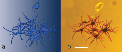
Confocal laser scanning microscopy (CLSM) is an analytical technique usually applied to biological and medical samples. It is used to produce high resolution in-focus three dimensional images of thick sections by targeted fluorescence. Trichomes held in amber fluoresce in the far red range whereas amber fluoresces in the ultraviolet. This allows the trichomes to be resolved easily from the amber by CLSM. Samples of amber from two regions were selected for analysis. Baltic amber (Eocene) is well known for its trichome inclusions which have, in the past, been used as a diagnostic feature of that amber. Mexican amber (Middle Miocene) from Simijovel, Chiapas, Mexico also contains abundant trichomes. Samples of amber from both these locations were successfully imaged and re-constructed in 3D using CLSM. This technique enables detailed analysis of the trichome structure without damaging the sample.

RESUMO [in Portuguese]

A microscopia confocal de scanning laser é uma técnica analítica que é geralmente aplicada em amostras biológicas e médicas. Esta técnica produz imagens tridimensionais em foco de alta resolução de secções espessas por fluorescência. Os tricomas aprisionados no âmbar fluorescem na zona mais distante do espectro visível do vermelho enquanto que o âmbar fluoresce no ultravioleta. Isto permite identificar facilmente os tricomas do âmbar por microscopia confocal de scanning laser. Amostras de âmbar nas duas regiões foram seleccionadas para análise. O âmbar do Báltico (Eocénico) é conhecido pelas suas inclusões de tricomas que foram já usadas como características diagnóstico para este tipo de âmbar. O âmbar do México (Miocénico médio) de Simijovel, Chiapas, México também contém tricomas em abundância. Amostras destas duas localizações foram reconstruídas com sucesso em 3D usando microscopia confocal de scanning laser. Esta técnica permite efectuar análises detalhadas das estruturas dos tricomas sem prejuízo da amostra.

INTRODUCTION

The use of confocal laser scanning microscopy (CLSM) for the study of inclusions in amber is a recent development. Böker & Brocksch (2002) produced a series of images of insects in Baltic amber using CLSM and identified the potential for 3D imaging of minute detail of taxonomically important morphological structures such as the mandibles and genital organs. Since then, however, very little has been published on CLSM analysis of amber inclusions despite the apparent benefits.

Another study using this technique, amongst other microscopic techniques, looked at some Spanish amber from Álava (Ascaso et al. 2003, 2005). The study looked at a protozoan with fungal hyphae trapped in amber and produced a 3D image based on a series of optical sections recorded by the CLSM. It is perhaps surprising, considering the detail and quality of the images that can be produced, that more studies have not incorporated this technique.  More recently, Speranza et al. (in press) have used light microscopy, CLSM & widefield fluorescence microscopy to image microscopic fungi embedded in amber, thus confirming the benefits of a ‘fluorescence’ approach. 

Trichomes are present in the vast majority of angiosperms and have been considered for some time to be of importance in comparative systematic studies (Theobald et al.  1979). They have an important role as defensive structures, especially in repelling phytophagous insects (Levin 1973). There is often more than one trichome type on any one taxon, but certain types may be more common to one taxon than another (Theobald et al. 1979). The taxonomic value of trichomes is therefore limited, but the Baltic trichomes are not inconsistent with them being from a type of oak.  

In this study, certain elements of the microflora are examined. The “stellate hairs”, or trichomes, are common in Baltic amber and have been considered as a characteristic of this type of amber (Weitschat & Wichard 2002). These trichomes are found associated with, and attached to, the male oak flowers and are therefore thought to belong to oak also (Weitschat & Wichard 2002) although there may be more than one type of trichome present. During this study samples from Chiapas, Mexico were also examined and were found to contain abundant trichomes. No previous record of trichomes in Mexican amber has been found in the literature.

Weitschat and Wichard (2002) describe the ‘stellate hairs’ found in Baltic amber as structures that develop on the flower and leaf buds of  the oak that are shed in great numbers every year. They also state that no studies have been able to clarify the origin or their significance for amber.

The interpretation of what the inclusions in Baltic amber represent in terms of their ecology has been the subject of much debate as many of the insect inclusions suggest a wide range of biotopes (Weitschat and Wichard 2002). Even individual pieces of amber seem to contain species from what would currently be both temperate and tropical regions (Weitschat 1997). This may suggest that species represent within the amber could have had a wider range then than their extant relatives (Weitschat and Wichard 2002).

In the present study we present a technique which uses CLSM and 3D imaging to analyse the structure of trichomes.

JPT No. 7 – The Setup, Use And Efficacy Of Sodium Polytungstate Separation Methodology With Repsect To Microvertebrate Remains

Jonathan S. Mitchell1,2 and Andrew B. Heckert1

1- Department of Geology, Appalachian State University, 572 Rivers Street, Boone, North Carolina, USA

2- Committee on Evolutionary Biology, University of Chicago, 5801 South Ellis Avenue, Chicago, IL 60637-1546; mitchelljs@uchicago.edu (current address)

ABSTRACT 

Concentrated deposits of small remains from vertebrates, termed microvertebrate sites or vertebrate microsites, are a unique and detailed source of information about the history of life. Collecting fossils from these sites, however, presents unique challenges. The most time consuming, and thus most deterring, aspect by far is the separation of the fossils from the sediment. This study attempts to quantify to what extent the use of sodium polytungstate (=sodium metatungstate, Na6H2W12O40, abbr. SPT) filtration increases fossil concentration, how quickly fossils sink in SPT solutions, and what is a good working density for SPT.  We do this by generally following the methodology set out by previous authors, although with some substantial modifications, on an Upper Triassic deposit dominated by clay minerals and lithic fragments, as well as on a second, smaller quartz sand dominated microsite. We also provide a revised and detailed guide with our modifications to former practices and our recommendations to other workers interested in creating a SPT laboratory, including the strong advisory to work over thin plastic sheets, as SPT can react with metal and adheres strongly to glass when it crystallizes.

Our experiments have shown a significant improvement in fossil concentration (from ~2% of the clasts being fossils to ~19%) at the main site, with a sample from the other site showing the treated concentrate as 25% fossil. We have also found very few fossils in the float (<0.5%), but noticeable rates of fossil loss in SPT solutions above ~2.80 g/mL (up to 16%).  Further, we have found that 2.75 g/mL is a good working density for several lithologies, as it is high enough to float most rock, low enough to sink most fossils, and low enough to be manageably maintained. SPT has, in processing one particularly rich site, saved many person-hours that otherwise would have been spent picking through less concentrated sediment.

INTRODUCTION

Studies of microvertebrate fossils (or vertebrate microremains) are becoming increasingly common (Sankey and Baszio, 2008).  Despite providing a wealth of information about past environments and ecosystems, microvertebrate studies are stymied by the difficulty of collection. The process of collecting and isolating large numbers of what are, by definition, tiny and potentially fragile fossils can be extremely time consuming and tedious. Methods have been developed to expedite this process (Cifelli et al 1996:18, Wilborn 2009), yet the fundamental methodology remains extremely similar to its original construction (Hibbard 1949). Further, no one to date has quantified the efficacy of these methods for vertebrate paleontology (though see Bolch, 1997 for dinoflagellates, Krukowski, 1988:315 for conodonts, Murray and Johnston, 1987:319 for heavy minerals in sediments, and Munsterman and Kerstholt, 1996 for palynological experiments).  After a site has been located, it is typically surface collected, then excavated, with vast quantities of sediment being taken away. These bags of sediment are then screen-washed in an attempt to remove as much clay and fine silt, while simultaneously retaining as many fossils, as possible.  After screen-washing, there typically remains a significant volume of concentrate, which is usually composed primarily of non-fossil clasts. After this step, a researcher, preparator or volunteer must go through the screen-washed concentrate one pinch of sediment at a time under a light microscope, isolating and removing individual fossils. These standard techniques for recouping micro-vertebrate  remains from concentrate are extremely time intensive and often dependent on an extensive time investment by students or volunteers (Hibbard, 1949, Grady 1979).

Inevitably, there will be fossiliferous concentrate that needs to be hand picked. The advantage of heavy liquid separation techniques is that they reduce the amount of unnecessary (nonfossiliferous) sediment that needs to be picked through. Traditionally heavy liquid separation was often accomplished using bromide liquids, with their extremely toxic nature representing a significant drawback (Cifelli et al., 1996:17, Murray and Johnston, 1987:317, Murry and Lezak, 1977:17). Murray and Johnston (1987:319) compared SPT to tetrabromoethane (TBE) and found no significant difference for sedimentological applications in the final product, noting only cost and viscosity (concurrent with Cifelli et al., 1996:17-18, though see Jeppsson and Anehus, 1999:57 and below for explanations of this discrepancy) as drawbacks to SPT.

Heavy liquid concentration, regardless of the chemicals used, makes picking both easier and more enjoyable (finding lots of fossils instead of few fossils per unit volume). This also maximizes research time by speeding up fossil recovery. The heavy liquid discussed here, sodium polytungstate (=sodium metatungstate, Na6H2W12O40, abbr. SPT) can be purchased dry and dissolved in deionized water to any desired density from 2.00 g/mL to 3.10 g/mL.

Tungsten compounds have been found to be safe in general (Kazantzis,1979), and sodium polytungstate, unlike bromides and kerosene, is generally regarded as safe unless ingested or applied to the eye (Cifelli et al., 1996:17 and many references therein, also see the Material Safety Data Sheet [MSDS, linked in references] or equivalent safety documentation). Further, sodium polytungstate can be reused continually, assuming it is taken care of properly. It is however, quite expensive (>$200 per 0.1kg), and traditionally difficult to obtain (though the Internet has reduced that problem, as a simple Google® search will reveal). Further, we followed the recommendations of Callahan (1987:765) in using bleached coffee filters instead of filter paper (contra Murray and Johnston, 1987:318) as they appear to speed recovery, but they also seem to have allowed clays to enter and discolor the SPT (though no other side effects have been confirmed, they may have absorbed some of the SPT as a precipitate, see McCarty and Congleton, 1994:198). Six et al. (1999) describe a process of cleansing SPT of organic contents by percolation through a column of activated carbon, and similar methods may work for the removal of clay, though we did not test this, and Murray and Johnston (1987:317-319) and Callahan (1987:765) both argue that laboratory-grade filter paper is enough. Yet as a possible (though unlikely) consequence of clay contamination (clay from a previously separated site contaminating future sites’ fossils) we advise caution in performing geochemical analyses on SPT separated fossils without heavily rinsing them until further studies on the solution’s effects and the efficacy of clay removal are performed.

Despite these modest drawbacks, SPT still provides a powerful tool for the paleontologist/preparator’s arsenal, as we found it easy to use, efficient, and very effective (see below). The ability to continually reuse it, as well as its speed and efficacy, make it cost effective in the long run, albeit a rather large initial investment is required. Here we outline the materials we recommend for a sodium polytungstate separation laboratory, the methods of separation, and the efficacy of the system.
JPT No. 6 – Abstract Book for the International Conference on Geological Collection and Museums: Mission and Management

NOTA DE ABERTURA

O presente livro reúne os resumos das contribuições dos participantes na Conferência Internacional Colecções e Museus de Geociências: missão e gestão, organizada pelo Centro de Estudos de História e Filosofia da Ciência e pelo Museu Mineralógico e Geológico, secção do Museu de História Natural) da Universidade de Coimbra (Museu da Ciência), agrupados por secções temáticas, de acordo com a sua apresentação. Os grandes desafios desta conferência internacional foram situados em torno das questões da missão e da gestão das colecções de objectos geológicos actualmente repartidas por um significativo número de instituições públicas e privadas, de que se destacam sobretudo universidades, laboratórios do Estado, museus e associações de carácter cultural e/ou científico. Nestes domínios, registamos com grande apreço a resposta da comunidade científica envolvida com este domínio tão particular do nosso património científico e cultural, sublinhando a espontaneidade e diversidade de propostas de intervenção surgidas. É nossa firme convicção que a apresentação e discussão de exemplos de boas práticas, de ideias de trabalho e de linhas orientadoras e políticas de gestão das colecções, será enriquecedora e poderá reflectir-se de forma positiva na adopção de estratégias de facilitação do acesso à plena fruição, científica e cultural, deste importante património, independentemente da sua natureza, localização geográfica ou tutela. Sublinhamos também a presença de um significativo lote de comunicações alinhadas pelos novos paradigmas de estudo, valorização e apresentação do património geológico in situ, rumos de actividade de grande potencial cultural e mesmo económico. Finalmente, manifestamos o nosso desejo que este primeiro encontro de profissionais envolvidos com a comunicação e divulgação museológica das Geociências se venha a constituir num fórum permanente de discussão e troca de experiências que, apoiado em estruturas informais ou outras, tendo em vista o constante aperfeiçoamento destas actividades-chave para a formação e divulgação de uma cultura geocientífica dos nossos cidadãos.

José M. Brandão Comissão Organizadora

JPT No. 5 – Preparation techniques applied to a stegosaurian dinosaur from Portugal

Ricardo Araújo1,2, Octávio Mateus1,2, Aart Walen3 and Nicolai Christiansen1,3

1- Museu da Lourinhã, Rua João Luís de Moura, 2530-157 Lourinhã. ricardo_araujo@museulourinha.org ; omateus@museulourinha.org

2- CICEGe, Faculdade de Ciências e Tecnologia, Universidade Nova de Lisboa, , 2829-516 Caparica, Portugal.

3- Creatures and Features, Rijndijk 17, NL-6686 MN, Doornenburg, Netherlands. cxfaartwalen@gmail.com ; cxfnac@gmail.com

ABSTRACT 

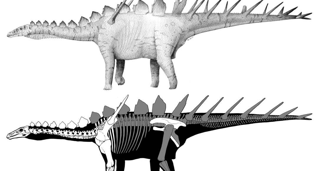
General vertebrate paleontological techniques that have been used in the Museum of Lourinhã (Portugal) are presented here, in particular those applied to a stegosaurian dinosaur skeleton, Miragaia longicollum. A monolith jacket technique using polyurethane foam and plaster is presented. Mechanical preparation techniques combining the use of an electric grinder and airscribes proved effective during the initial phases of preparation on well-preserved bone embedded in hard matrix. We also present a technique to mould monoliths in the early stages of preparation, creating a thin silicone rubber mould in several contiguous parts. To mould and cast monoliths before removing individual bones has proven valuable for the preservation of taphonomic data and for display purposes. Polyurethane resin combined with plaster is useful for small casts, while polyester resin applied in four layers is the preferred technique for larger casts. The four layers are composed of: a first thin layer of polyester resin with bone colour; followed by another layer of polyester resin of sediment colour and containing glass microspheres to make it thicker. The third layer is composed of fibre glass chopped strands, and the fourth is composed of fibre glass mats embedded in plain polyester resin.  3D scanning and digitization techniques where tested for the storage of osteological information of individual bones and proved very promising.

INTRODUCTION

This paper describes the excavation, preparation, moulding, casting, and 3D scanning of a Late Jurassic dinosaur from Lourinhã, Portugal. This area is rich in dinosaurs and other vertebrates (see Antunes and Mateus 2003, Mateus 2006, and references therein). Fossil collecting and preparation in Museum of Lourinhã commenced in 1984 when the first specimens of crocodiles and dinosaurs were collected. Initially, the equipment was rudimentary little more than chisel and hammer was used. Out of curiosity, the first electric (air)scribe was acquired in 1998. The increasing number of fossils collected also brought awareness of the vital importance of preparation in the paleontological collections. The first full time preparator – Dennis Roessler – was then hired in 2001. The subsequent work was then developed by Nicolai Christiansen, Ricardo Araújo, Rui Lino and Alexandra Tomás, in consultation with Aart Walen and Museum research scientists.

Currently, Museum of Lourinhã has a modern laboratory making use of techniques designed on the basis of other preparation laboratories and adapted to our own conditions (fig. 1). “The preparation techniques applied in the Museum of Lourinhã are presented here using, as a study case, the entire process of preparation of the stegosaurian dinosaur specimen ML433 (fig. 2). This specimen was discovered in 2001 by Rui Soares, who initially discovered an exposed osteoderm (fig. 2) and formally described as a new taxon: Miragaia longicollum (Mateus et al. 2009).

JPT No. 4 – A quick method for collecting modern small-scale ichnological and sedimentological structures

Thomas A. Stidham1 and Jane Mason2

1  Department of Biology, Texas A&M University, 3258 TAMU, College Station, Texas 77843-3258, USA. e-mail: furcula@mail.bio.tamu.edu

2  Division of Vertebrate Paleontology, Florida Museum of Natural History, Dickinson Hall, University of Florida, Gainesville, Florida 32611-7800, USA e-mail: jmason@flmnh.ufl.edu

ABSTRACT 

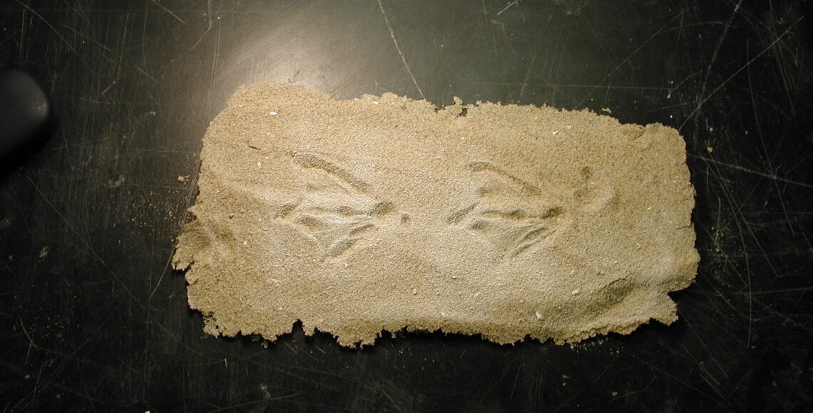
We describe a method for collecting recent superficial sedimentary structures, such as ripples, tool marks, and trackways. The surface first is consolidated using one of a variety of materials (acrylic, Butvar, or dust hardener), and then reinforced using cellulose acetate and collected. Sediment grains and areas of sediment surface can be excised after collection and cleaned for detailed study. This method is useful on a variety of sediment sizes, and it is inexpensive and relatively quick to implement.

INTRODUCTION

Modern trackways and other ichnological traces are fundamental tools used to research fossil tracks and sedimentary structures, including ripples and tool marks. However, commonly used methods of collecting modern footprints include the laborious use of plaster or latex (Goodwin and Chaney, 1994). Both of these methods are inadequate to accurately record traces on unconsolidated substrates without crushing the print or otherwise distorting the trace or structure. In addition, neither of these methods consistently and accurately records details of the substrate’s sedimentology, including grain size and sorting. These features of the sediment may influence the formation of an ichnological trace or sedimentary structure. In order to circumvent some of the problems with these other methods, we have developed a technique that records small-scale surficial sedimentary structures with little, if any, distortion, requiring less labor, and preserving more data. It is similar to the use of acetate molds in other areas of paleontology (Darrah, 1936; Rigby and Clark, 1965).

JPT No. 3 – Stone-splitters and expansive demolition agents in vertebrate paleontological excavations

Mateus, O. and Araújo, R.

CICEGe, Faculdade de Ciências e Tecnologia, Universidade Nova de Lisboa, Quinta da Torre, P-2829-516 Caparica, PORTUGAL; and Museu da Lourinhã, Rua João Luis de Moura, 2530-157 Lourinhã, Portugal; omateus@fct.unl.pt and paradoxides@gmail.com

ABSTRACT 

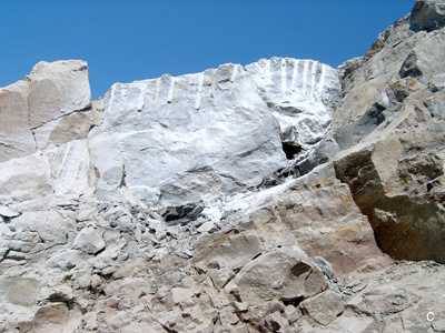
Two techniques (stone-splitters and demolition agents) are revealed to be efficient methods for breaking large stone blocks in extreme paleontological excavation. In certain conditions – where security factors, permission issues, terrain conditions, rock properties are problematic – the traditional methods for breaking large rock blocks cannot be applied (e.g. crane trucks or explosives).  Using an expansive demolition agent or stone-splitters after drilling equidistant holes not only allows a cheap, quick and safe solution but also permits precise removal of up to 9 ton blocks.

Stone-splitters are a three-part tool that when inserted linearly and equidistantly along a brittle rock mass cause a precise fracture.

INTRODUCTION

Some paleontological excavations require the removal of massive overburden composed of hard rock bodies. Collecting in such conditions often requires the use of explosives (that could damage the fossils) or heavy machinery. Moreover, explosives and heavy machinery (e.g. bulldozers, crane trucks) may not be a solution due to security factors, permission issues, terrain conditions, and rock properties. On the one hand, crane trucks are not always able to access the fossil sites and explosives are also avoided due to high accident risk, the need of specific training, requirement of formal permissions, and the danger of damaging the fossils. Due to several constraints, some rocks and layers have to be removed by hand, using a pneumatic or electric jackhammer, which is time-consuming, causes rapid equipment deterioration, and is physically exhausting.